Saturday, May 17, 2014

REPOST: 7 hand exercises to ease arthritis pain

Arthritis pain can be debilitating, but that doesn’t mean you can’t do anything about it. This article gives instructions for exercises that help manage the pain in arthritic hands.

http://www.healthline.com/health-slideshow/arthritis-hand-exercises#1
Image Source: healthline.com

Painful Hands

Arthritis wears away at the cartilage and synovial lining of a joint, which is the cushioning material between bones. When arthritis affects the joints of the hands, it can cause pain and stiffness. That pain can get worse whenever you use the hand a lot—for example, when typing on a computer keyboard or gripping utensils in the kitchen. You may also lose strength in your hands. Weakness in your hands can make it hard to do even the simplest everyday tasks, such as opening jars.

Treating Hand Arthritis

There are some medicinal options for treating hand arthritis. You can take pain-relieving medicines by mouth. There are also injections of steroid medicines to decrease swelling in the joints, and splinting to support and protect your hands. If these options don’t work, you may need to have surgery to fix the damaged joint.
As far as home treatments: one easy and noninvasive way to keep the joints flexible, improve range of motion, and relieve arthritis pain is by doing hand exercises.

Exercise #1: Make a Fist

You can do this easy exercise anywhere, and any time your hand feels stiff. Start by holding your left hand up straight. Then, slowly bend your hand into a fist, placing your thumb on the outside of your hand. Be gentle—don’t squeeze your hand. Open your hand back up until your fingers are straight once again. Do the exercise 10 times with the left hand. Then repeat the whole sequence with the right hand.

Exercise #2: Finger Bends

Start in the same position as in the last exercise, with your left hand held up straight. Bend your thumb down toward your palm. Hold it for a couple of seconds. Straighten your thumb back up. Then bend your index finger down toward your palm. Hold it for a couple of seconds. Then straighten it. Repeat with each finger on the left hand. Then repeat the entire sequence on the right hand.

Exercise #3: Thumb Bend

First, hold your left hand up straight. Then, bend your thumb inward toward your palm. Stretch for the bottom of your pinky finger with your thumb. If you can’t reach your pinky, don’t worry. Just stretch your thumb as far as you can. Hold the position for a second or two, and then return your thumb to the starting position. Repeat 10 times. Then do the exercise with your right hand.

Exercise #4: Make an “O”

Start with your left hand pointing straight up. Then, curve all of your fingers inward until they touch. Your fingers should form the shape of an “O.” Hold this position for a few seconds. Then straighten your fingers again. Repeat this exercise a few times a day on each hand. You can do this stretch whenever your hands feel achy or stiff.

Exercise #5: Table Bend

Place the pinky-side edge of your left hand on a table, with your thumb pointed up. Holding your thumb in the same position, bend the other four fingers inward until your hand makes an “L” shape. Hold it for a couple of seconds, and then straighten your fingers to move them back into the starting position. Repeat 10 times, and then do the same sequence on the right hand.

Exercise #6: Finger Lift

Place your left hand flat on a table, palm down. Starting with your thumb, lift each finger slowly off the table—one at a time. Hold each finger for a second or two, and then lower it. Do the same exercise with every finger of the left hand. After you’re done with the left hand, repeat the entire sequence on the right hand.

Exercise #7: Wrist Stretch

Don’t forget about your wrists, which can also get sore and stiff from arthritis. To exercise your wrist, hold your right arm out with the palm facing down. With your left hand, gently press down on the right hand until you feel a stretch in your wrist and arm. Hold the position for a few seconds. Repeat 10 times. Then, do the entire sequence with the left hand
 
I’m Brittany Perskin, and I’m a licensed physical therapist, health nut, and hand therapy enthusiast. If you want to read some more of my thoughts and updates, follow this Google+ page.

Monday, March 24, 2014

REPOST: Watch this Parkour video if you want the experience but not the broken bones

Anyone who watches this video will surely have a glimpse of what it feels like to do parkour. This post from USA Today features James Kingston of parkour group Ampisound with a camera fixed on his head while he performed the extreme sport. 

                                                 
How is this not a video game yet?

James Kingston is a British member of the Parkour group Ampisound. In this video, he fixed a camera onto his head and filmed himself jumping and leaping around Cambridge, England like some sort of flying squirrel.
parkour1
Image Source: usatoday.com
My favorite part about the video is that they didn’t use some dubstep-bass-drop insanity that every other extreme sport video uses now, and for that, I applaud them.

parkour2
Image Source: usatoday.com
Also, does this guy have any cartilage in his knees at this point? This can’t be good for the joints.
Hi! I’m Brittany Perskin. I am a physical therapist who enjoys extreme sports, particularly parkour. Follow me on Twitter for more related stories.

Tuesday, January 21, 2014

REPOST: For Young Athletes, Injuries Need Special Care

Good news for young athletes! Hospitals in the US now provide more sports medicine programs that are tailored for kids. According to The Wall Street Journal, the new surgical techniques and physical therapy protocols aim to protect the growing bones of young patients and to provide them with encouragement and support, especially if they are "upset to be sitting out of a beloved sport." Read the full report below. 

(Kaylyn Lambertt, a high-school soccer player and now a freshman at Florida State University, had surgery on her left hip her junior year and her right hip her senior year. Leslie Russell) Image Source: online.wsj.com

Children's hospitals are expanding programs to care for a fast-growing category of young patients: injured athletes.

The rehabilitation needs of children and teens are different than those of adults. More sports medicine programs are working exclusively with young athletes, using surgical techniques and physical therapy protocols that don't interfere with growing bones and cartilage.

One aim of this is to prevent affecting the growth plate—the area of growing tissue near the end of long bones in children and teens. For example, while adults may lift heavier weights to build muscle during physical therapy, pediatric patients may do higher repetitions with lower resistance to avoid hurting growing bones, muscles and tendons. The programs also offer encouragement and support for kids upset to be sitting out of a beloved sport.

More than 3.5 million children a year receive treatment for sports injury, according to Stop Sports Injuries, a campaign whose backers include the American Orthopaedic Society for Sports Medicine. And high-school athletes account for an estimated 2 million sports injuries each year. While concussions account for about 15% of youth sports injuries, experts say many sports carry risks for musculoskeletal injuries, in large part due to increased emphasis on year-round competition, single-sport concentration and intense training regimens.

A study published last year by Boston Children's Hospital warned that children of all ages are sustaining significant sports injuries that require surgical intervention. "In the past we'd put a cast on a broken leg, take it off six to 13 weeks later and send kids home," says Lyle Micheli, director of the hospital's division of sports medicine. "Now we realize we have to very systematically rehabilitate these kids for strength and basic function, and determine when it is safe for them to return to play."


Image Source: online.wsj.com

While injuries from recreational activities such as biking have fallen over the last decade, team sports including football and soccer saw injuries rise by 22.8% and 10.8% respectively, according to a study last year by Cincinnati Children's Hospital Medical Center.

Doctors are seeing more overuse injuries. There has been a fivefold increase since 2000 in the number of shoulder and elbow injuries among youth baseball and softball players, according to Stop Sports Injuries.

Children's Hospital & Research Center Oakland, in California, last fall opened a Sports Medicine Center for Young Athletes at its Walnut Creek campus. "It's hard for kids to do rehabilitation next to an 85-year-old stroke victim or a 75-year-old cancer patient," says Nirav Pandya, the center's director, and an orthopedic surgeon. The center and many other pediatric clinics offer classes and programs to help kids improve sports performance while avoiding injury.

Physical therapy after injury and surgery, such as repair to the anterior cruciate ligament in the knee, is covered by insurance for varying periods. After that, clinics may design a regimen children can perform at home or at a local fitness facility.

Jeremy Frank, a pediatric orthopedic surgeon at Memorial Healthcare System's Joe DiMaggio Children's Hospital in Hollywood, Fla., says that there is often little pain a week after minimally invasive ACL surgery, so young people "think they are good to go and don't realize they have six months of rehabilitation in front of them." Often, he says, there is a "bargaining moment" where his young patients try to get him to approve more activity than they are ready for. Patients are generally referred to Memorial's two U-18—for Under 18—physical rehabilitation clinics in Coral Springs, Fla., where therapists work with families and coaches to stress the importance of healing.


Dr. Frank, U-18's assistant director, says while the vast majority of athletes get back to sports and do well, there are times when a young patient sustains multiple injuries such as a third ACL tear. "You have arthritic changes in your knee, and you have to stop playing soccer," he says.

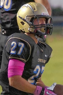
Dylan Rupert, 17, a running back and captain of the Cypress Bay High School football team in Weston, Fla., tore his ACL during play last fall. His parents opted for a repair technique, which surgeons are more often using in pediatric patients. The procedure avoids drilling through the growth plate and may decrease risks of future pain and re-injury. The surgery used part of Dylan's own hamstring rather than a cadaver tissue more commonly used in adults. He started rehab at Coral Springs three days after his Oct. 22 surgery.

The injury was devastating for Dylan. It came just as he was getting the attention of college coaches, says his mother, Monica Puga-Finch, an information technology program director at the clinic's parent Memorial. In his first physical therapy session, senior therapist Whitney Chambers helped calm his fears, but "told him that she was going to push him, and he couldn't say 'I can't.' " As the sessions continued twice a week he would often come out sweating and sore but excited, "with a sense of accomplishment," Ms. Puga-Finch says. Ms. Chambers helped with the emotional aspects of being sidelined, encouraging him to go to practices and games with his team. His rehabilitation is expected to take six to eight months. He plans to return to sports in college.

Ms. Chambers says physical therapy after the growth-plate sparing procedure is more conservative than for the traditional ACL reconstruction. It starts with protective weight bearing exercises using crutches and a knee brace, gentle range of motion work, and ice and electrical stimulation for swelling and pain control. Then she works on strengthening muscles and restoring joint flexibility. To make it more fun, she uses games or obstacle courses.

The clinic uses screening questionnaires to identify kids at risk of depression, who may be referred to a child psychologist.

Kaylyn Lambertt who has played soccer from the age of 6, was a junior in high school when she felt a searing pain in her left hip during a game in December 2010. She continued to play for months as it got worse. Her labrum, part of her hip joint, was torn in two places, with a socket out of place. A lump on her bone was wearing down the cartilage every time she walked or ran. She had surgery to repair the damage in 2011, followed by months of rehabilitation with Ms. Chambers.

She returned to soccer her senior year, but began feeling pain, this time in her right hip. Dr. Frank told her that she had torn the labrum. She underwent a second surgery in December 2012. She returned to Ms. Chambers and realized during their talks that "soccer isn't everything." Now a freshman at Florida State University she plays a pickup game of soccer now and then, but is focused on what Ms. Chambers inspired her to chose as a career: physical therapy.

(Dylan Rupert, 17, of Weston, Fla., had surgery to repair a torn ACL last fall. I Three Photography) Image Source: online.wsj.com


Brittany Perskin here, your child-friendly physical therapist in Los Angeles, CA. Follow me on Twitter for more updates on physical therapy and sports medicine.

Thursday, December 19, 2013

REPOST: The Ultimate Beginner’s Guide to Parkour

Brett and Kate McKay share in The Art of Manliness an exhaustive introduction to parkour, the sport “where you jump from buildings and vault over walls.”
Image source: artofmanliness.com

You’ve seen it on TV shows such as American Ninja Warrior (and not so seriously in The Office) as well as in movies like Casino Royale and The Bourne Ultimatum. If you’ve played Assassins Creed or Mirror’s Edge, you’ve even done it, virtually, at least.

I’m talking about parkour.


Video source: youtube.com

Yeah. That sport where you jump from buildings and vault over walls. Many men are drawn to parkour even if they’re not entirely sure what it is. It’s captivating to see someone move through an environment in ways we had previously not conceived of, and inspiring to witness the human body pushing the very limits of its capabilities. Plus, it just looks like so much fun and it seems like an important skill to have during the zombie apocalypse when you’ll need to be able outrun a pack of vicious brain-eaters (depending on your theory of their bipedal capabilities, of course).

To learn more about parkour I visited the Tempest Freerunning Academy in Los Angeles to talk to parkour/freerunning instructor, stuntman, Ninja Warrior veteran, and epic handlebar mustache owner Brian Orosco .

What is Parkour?

Video source: youtube.com

Parkour is all about moving through your environment efficiently and naturally. Parkour practioners, who are often called traceuers (from the French for ‘to trace’), jump, climb, and vault over obstacles in their path. Their goal is to get from point A to point B as efficiently as possible.

The history of parkour is actually pretty fascinating. It got its start in France and has its roots in military escape and evasion tactics and 19th century physical culture. In fact, the word “parkour” originates from the French phrase “parcours du combattant:” the obstacle course-based method of training used by the French military. So while we think of parkour today as simply an interesting form of recreation, it was actually developed as a tactical skill and way to build the fitness of soldiers.

The Difference Between Parkour and Freerunning
Image source: artofmanliness.com


Parkour and freerunning get used interchangeably. While they share a lot in common, there is a small difference.

Parkour is simply about maneuvering through your environment efficiently using jumps, swings, and vaults. No need for flips, wall spins, and other acrobatics. With freerunning, efficiency is less of a concern, and you can throw in these types of cool-looking acrobatic movements as well.

So when you’re watching YouTube videos of people doing flips and spins off walls, that’s freerunning; if they’re just jumping and vaulting over urban obstacles without acrobatics, they’re doing parkour.


Read the continuation of this article here for more on the basics of parkour.

Have you tried parkour? If yes, then let me know what you think of this urban workout. Add me, Brittany Perskin, on Facebook and let’s trade stories about this exciting sport!

Friday, November 15, 2013

REPOST: Frankfort hand therapist boasts rare certification

Hand therapy is a highly specialized form of therapy conducted by physical therapists on those with injuries or conditions affecting the hands. This report from The Herald-News.com delves more into the subject through the experiences of Frankfort, Illinois-based hand therapist, Rick Brasel. 


Certified Hand Therapist Rick Brasel works the muscles of Anthony Catezone of Frankfort who is recovering from a broken elbow. | Susan DeMar Lafferty~IMAGE SOURCE: Sun-Times Media

Rick Brasel’s work station is filled with so many toys and devices that one of his clients refers to him as “Inspector Gadget.”

There’s bright-colored putty, tiny manipulatives, wheels, balls and grippers — all of which he shares with those who visit him.

On a recent afternoon, Nick Rizzo was manipulating the manipulatives with his fingers, squeezing the gripper and lifting a heavy blue ball. Randy Collette wrestled with some very tough putty, while Anthony Catezone rolled his arm back and forth with a wheel.

It’s all in a day’s therapy.

Brasel, an occupational therapist at Flexeon Rehabilitation in Frankfort, recently became a certified hand therapist, one of an elite group of 5,800 hand specialists worldwide — a “gold standard” among therapists, he said.
His gadgets are intended to help his clients regain full use of their hands and arms.

What makes the hand so special?

“Ultimately, it’s everything we do. The hand is most important in terms of overall function,” Brasel said, admitting he’s biased. Hands are required for doing basic activities, such as eating, brushing teeth and combing hair.
“A large portion of our body does nothing during the day. But hands never get a break. Hands can do so many things. If you cannot see, you rely on feeling. You lose a lot of functions that you take for granted if you can’t use your hands,” he said.

Just ask his clients.

Rizzo suffered a stroke a few months ago and lost all functions on his right side. He was unable to fasten his shirt buttons, tie his shoes or hold a coffee cup. After a few weeks with Brasel, “I can tie my shoes and dress myself and do a lot of things I couldn’t do before,” Rizzo said.

Brasel works Rizzo’s hand and arm muscles in a variety of ways to help him regain strength, endurance, coordination and sensation.

Read the entire article to learn about Rick Brasel's hand therapy practice here.

Brittany Perskin is a physical therapist who hopes to someday specialize in hand therapy. Learn more about physical therapy and wellness by following updates in this Twitter account.

Monday, October 21, 2013

REPOST: Death-Defying Parkour Stunts Will Have You On Edge

Alexander Rusinov, a Russian gymnast and freerunner, attempts one of the most death-defying stunts choreographed in the world of parkour. Watch him perform his stunts on this article from The Huffington Post.


"Do not try this without training," the video warns.

After watching the death-defying parkour stunts, we can guarantee we won't.

Russian gymnast and freerunner, Alexander Rusinov, tempts fate on some of the highest buildings he can find. While Alexander has had some very specialized training, not even his incredible strength can make those high-flying acts look easy.

Alexander's translated video description reads:
“Once again I have overcome my fears. I have taken my victory over these wildly conceived ideas, and have brought them to life. This is my life and I have once again proven that I can trust in myself and my own strength.”
As much as this video made us nauseous, we secretly can't wait for the next one from Alexander and Dangerous Games 3.
Brittany Perskin is a physical therapist who loves doing parkour on her free time. More updates about her active lifestyle can be found on this Facebook page.

Thursday, September 19, 2013

REPOST: Nanotechnology In Medicine: Huge Potential, But What Are The Risks?

My colleagues and I recently had a discussion about nanotechnology. We talked about its the benefits and risks, and whether or not its advantages outweigh the uncertainties. The article below provides more details on nanotechnology.

Nanotechnology, the manipulation of matter at the atomic and molecular scale to create materials with remarkably varied and new properties, is a rapidly expanding area of research with huge potential in many sectors, ranging from healthcare to construction and electronics. In medicine, it promises to revolutionize drug delivery, gene therapy, diagnostics, and many areas of research, development and clinical application.

This article does not attempt to cover the whole field, but offers, by means of some examples, a few insights into how nanotechnology has the potential to change medicine, both in the research lab and clinically, while touching on some of the challenges and concerns that it raises.

What is Nanotechnology?

The prefix "nano" stems from the ancient Greek for "dwarf". In science it means one billionth (10 to the minus 9) of something, thus a nanometer (nm) is is one billionth of a meter, or 0.000000001 meters. A nanometer is about three to five atoms wide, or some 40,000 times smaller than the thickness of human hair. A virus is typically 100 nm in size.

The ability to manipulate structures and properties at the nanoscale in medicine is like having a sub-microscopic lab bench on which you can handle cell components, viruses or pieces of DNA, using a range of tiny tools, robots and tubes.
Image Source: medicalnewstoday.com

Manipulating DNA

Therapies that involve the manipulation of individual genes, or the molecular pathways that influence their expression, are increasingly being investigated as an option for treating diseases. One highly sought goal in this field is the ability to tailor treatments according to the genetic make-up of individual patients.

This creates a need for tools that help scientists experiment and develop such treatments.

Imagine, for example, being able to stretch out a section of DNA like a strand of spaghetti, so you can examine or operate on it, or building nanorobots that can "walk" and carry out repairs inside cell components. Nanotechnology is bringing that scientific dream closer to reality.

For instance, scientists at the Australian National University have managed to attach coated latex beads to the ends of modified DNA, and then using an "optical trap" comprising a focused beam of light to hold the beads in place, they have stretched out the DNA strand in order to study the interactions of specific binding proteins.

Nanobots and Nanostars

Meanwhile chemists at New York University (NYU) have created a nanoscale robot from DNA fragments that walks on two legs just 10 nm long. In a 2004 paper published in the journal Nano Letters, they describe how their "nanowalker", with the help of psoralen molecules attached to the ends of its feet, takes its first baby steps: two forward and two back.

One of the researchers, Ned Seeman, said he envisages it will be possible to create a molecule-scale production line, where you move a molecule along till the right location is reached, and a nanobot does a bit chemisty on it, rather like "spot-welding" on a car assembly line. Seeman's lab at NYU is also looking to use DNA nanotechnology to make a biochip computer, and to find out how biological molecules crystallize, an area that is currently fraught with challenges.

The work that Seeman and colleagues are doing is a good example of "biomimetics", where with nanotechnology they can imitate some of the biological processes in nature, such as the behavior of DNA, to engineer new methods and perhaps even improve them.

DNA-based nanobots are also being created to target cancer cells. For instance, researchers at Harvard Medical School in the US reported recently in Science how they made an "origami nanorobot" out of DNA to transport a molecular payload. The barrel-shaped nanobot can carry molecules containing instructions that make cells behave in a particular way. In their study, the team successfully demonstrates how it delivered molecules that trigger cell suicide in leukemia and lymphoma cells.

Nanobots made from other materials are also in development. For instance, gold is the material scientists at Northwestern University use to make "nanostars", simple, specialized, star-shaped nanoparticles that can deliver drugs directly to the nuclei of cancer cells. In a recent paper in the journal ACS Nano, they describe how drug-loaded nanostars behave like tiny hitchhikers, that after being attracted to an over-expressed protein on the surface of human cervical and ovarian cancer cells, deposit their payload right into the nuclei of those cells.

The researchers found giving their nanobot the shape of a star helped to overcome one of the challenges of using nanoparticles to deliver drugs: how to release the drugs precisely. They say the shape helps to concentrate the light pulses used to release the drugs precisely at the points of the star.

Nanofactories that Make Drugs In Situ

Scientists are discovering that protein-based drugs are very useful because they can be programmed to deliver specific signals to cells. But the problem with conventional delivery of such drugs is that the body breaks most of them down before they reach their destination.

But what if it were possible to produce such drugs in situ, right at the target site? Well, in a recent issue of Nano Letters, researchers at Massachusetts Institute of Technology (MIT) in the US show how it may be possible to do just that. In their proof of principle study, they demonstrate the feasibility of self-assembling "nanofactories" that make protein compounds, on demand, at target sites. So far they have tested the idea in mice, by creating nanoparticles programmed to produce either green fluorescent protein (GFP) or luciferase exposed to UV light.

The MIT team came up with the idea while trying to find a way to attack metastatic tumors, those that grow from cancer cells that have migrated from the original site to other parts of the body. Over 90% of cancer deaths are due to metastatic cancer. They are now working on nanoparticles that can synthesize potential cancer drugs, and also on other ways to switch them on.

Image Source: medicalnewstoday.com

Nanofibers

Nanofibers are fibers with diameters of less than 1,000 nm. Medical applications include special materials for wound dressings and surgical textiles, materials used in implants, tissue engineering and artificial organ components.

Nanofibers made of carbon also hold promise for medical imaging and precise scientific measurement tools. But there are huge challenges to overcome, one of the main ones being how to make them consistently of the correct size. Historically, this has been costly and time-consuming.

But last year, researchers from North Carolina State University, revealed how they had developed a new method for making carbon nanofibers of specific sizes. Writing in ACS Applied Materials & Interfaces in March 2011, they describe how they managed to grow carbon nanofibers uniform in diameter, by using nickel nanoparticles coated with a shell made of ligands, small organic molecules with functional parts that bond directly to metals.

Nickel nanoparticles are particularly interesting because at high temperatures they help grow carbon nanofibers. The researchers also found there was another benefit in using these nanoparticles, they could define where the nanofibers grew and by correct placement of the nanoparticles they could grow the nanofibers in a desired specific pattern: an important feature for useful nanoscale materials.

Lead is another substance that is finding use as a nanofiber, so much so that neurosurgeon-to-be Matthew MacEwan, who is studying at Washington University School of Medicine in St. Louis, started his own nanomedicine company aimed at revolutionizing the surgical mesh that is used in operating theatres worldwide.

The lead product is a synthetic polymer comprising individual strands of nanofibers, and was developed to repair brain and spinal cord injuries, but MacEwan thinks it could also be used to mend hernias, fistulas and other injuries.

Currently, the surgical meshes used to repair the protective membrane that covers the brain and spinal cord are made of thick and stiff material, which is difficult to work with. The lead nanofiber mesh is thinner, more flexible and more likely to integrate with the body's own tissues, says MacEwan. Every thread of the nanofiber mesh is thousands of times smaller than the diameter of a single cell. The idea is to use the nanofiber material not only to make operations easier for surgeons to carry out, but also so there are fewer post-op complications for patients, because it breaks down naturally over time.

Researchers at the Polytechnic Institute of New York University (NYU-Poly) have recently demonstrated a new way to make nanofibers out of proteins. Writing recently in the journal Advanced Functional Materials, the researchers say they came across their finding almost by chance: they were studying certain cylinder-shaped proteins derived from cartilage, when they noticed that in high concentrations, some of the proteins spontaneously came together and self-assembled into nanofibers.

They carried out further experiments, such as adding metal-recognizing amino acids and different metals, and found they could control fiber formation, alter its shape, and how it bound to small molecules. For instance, adding nickel transformed the fibers into clumped mats, which could be used to trigger the release of an attached drug molecule.

The researchers hope this new method will greatly improve the delivery of drugs to treat cancer, heart disorders and Alzheimer's disease. They can also see applications in regeneration of human tissue, bone and cartilage, and even as a way to develop tinier and more powerful microprocessors for use in computers and consumer electronics.

Image Source: medicalnewstoday.com

What of the Future and Concerns Surrounding Nanomaterials?

Recent years have seen an explosion in the number of studies showing the variety of medical applications of nanotechnology and nanomaterials. In this article we have glimpsed just a small cross-section of this vast field. However, across the range, there exist considerable challenges, the greatest of which appear to be how to scale up production of materials and tools, and how to bring down costs and timescales.

But another challenge is how to quickly secure public confidence that this rapidly expanding technology is safe. And so far, it is not clear whether that is being done.

There are those who suggest concerns about nanotechnology may be over-exaggerated. They point to the fact that just because a material is nanosized, it does not mean it is dangerous, indeed nanoparticles have been around since the Earth was born, occurring naturally in volcanic ash and sea-spray, for example. As byproducts of human activity, they have been present since the Stone Age, in smoke and soot.

Of attempts to investigate the safety of nanomaterials, the National Cancer Institute in the US says there are so many nanoparticles naturally present in the environment that they are "often at order-of-magnitude higher levels than the engineered particles being evaluated". In many respects, they point out, "most engineered nanoparticles are far less toxic than household cleaning products, insecticides used on family pets, and over-the-counter dandruff remedies," and that for instance, in their use as carriers of chemotherapeutics in cancer treatment, they are much less toxic than the drugs they carry.

It is perhaps more in the food sector that we have seen some of the greatest expansion of nanomaterials on a commercial level. Although the number of foods that contain nanomaterials is still small, it appears set to change over the next few years as the technology develops. Nanomaterials are already used to lower levels of fat and sugar without altering taste, or to improve packaging to keep food fresher for longer, or to tell consumers if the food is spoiled. They are also being used to increase the bioavailablity of nutrients (for instance in food supplements).

But, there are also concerned parties, who highlight that while the pace of research quickens, and the market for nanomaterials expands, it appears not enough is being done to discover their toxicological consequences.

This was the view of a science and technology committee of the House of Lords of the British Parliament, who in a recent report on nanotechnology and food, raise several concerns about nanomaterials and human health, particularly the risk posed by ingested nanomaterials.

For instance, one area that concerns the committee is the size and exceptional mobility of nanoparticles: they are small enough, if ingested, to penetrate cell membranes of the lining of the gut, with the potential to access the brain and other parts of the body, and even inside the nuclei of cells.

Another is the solubility and persistence of nanomaterials. What happens, for instance, to insoluble nanoparticles? If they can't be broken down and digested or degraded, is there a danger they will accumulate and damage organs? Nanomaterials comprising inorganic metal oxides and metals are thought to be the ones most likely to pose a risk in this area.

Also, because of their high surface area to mass ratio, nanoparticles are highly reactive, and may for instance, trigger as yet unknown chemical reactions, or by bonding with toxins, allow them to enter cells that they would otherwise have no access to.

For instance, with their large surface area, reactivity and electrical charge, nanomaterials create the conditions for what is described as "particle aggregation" due to physical forces and "particle agglomoration" due to chemical forces, so that individual nanoparticles come together to form larger ones. This may lead not only to dramatically larger particles, for instance in the gut and inside cells, but could also result in disaggregation of clumps of nanoparticles, which could radically alter their physicochemical properties and chemical reactivity.

"Such reversible phenomena add to the difficulty in understanding the behaviour and toxicology of nanomaterials," says the committee, whose overall conclusion is that neither Government nor the Research Councils are giving enough priority to researching the safety of nanotechnology, especially "considering the timescale within which products containing nanomaterials may be developed".

They recommend much more research is needed to "ensure that regulatory agencies can effectively assess the safety of products before they are allowed onto the market".

It would appear, therefore, whether actual or perceived, the potential risk that nanotechnology poses to human health must be investigated, and be seen to be investigated. Most nanomaterials, as the NCI suggests, will likely prove to be harmless.

But when a technology advances rapidly, knowledge and communication about its safety needs to keep pace in order for it to benefit, especially if it is also to secure public confidence. We only have to look at what happened, and to some extent is still happening, with genetically modified food to see how that can go badly wrong.

Hi, Brittany Perskin here. I work as a physical therapist in LA. Add me on Facebook and I’ll share with you the latest news on health, food, and wellness.